Ten years of painstaking work have led to the world’s first three-dimensional map of a fragment of the human brain with subcellular resolution. In one cubic millimeter of gray matter in the female brain, 57 thousand cells and 150 million synapses were found. The map revealed the details of all the intricacies of the nervous tissue in all the variety of cells found there, among which there were unknown to science, which brought the study of brain topology to a new level.
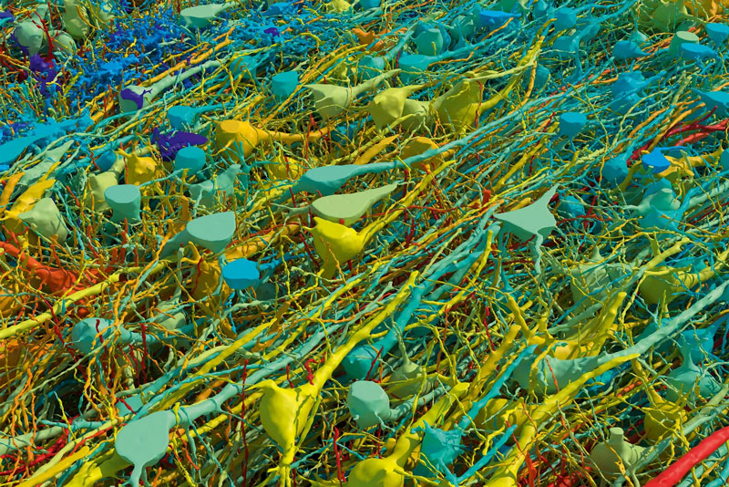
Image source: Harvard and Google
Back in 2014, a woman diagnosed with epilepsy underwent surgery to remove a tiny portion of the cortex cerebri – the gray matter tissue of the brain that covers both hemispheres with a thin layer. A group of scientists from Harvard University set out to restore as accurate a volumetric map of the sample as possible. To do this, the tissues were saturated with heavy metals, which bound to the lipid layer of the membranes of nerve cells and developed them for reading by an electron microscope. The tissues were then saturated with a hardening solution and cut into 34 nm thick slices, after which scanning and mapping began.
The volume of raw data for 2D slices was 1.4 PB. To process information on stitching images into one 3D object, scientists requested help from Google. The latter provided resources based on machine learning systems. In addition, scientists hand-painted fabrics in the most difficult cases and checked the accuracy of the model. For better visualization, the smallest cells were stained blue and the largest cells red. Cells of intermediate sizes received their colors from the usual spectral palette.
The result is the most detailed interactive map of the volume of human brain tissue with subcellular resolution to date. It is made publicly available so that any scientific team can use the map for their own research. The map revealed many surprising and even unexpected things. For example, scientists have discovered extensions of nerve cells in the form of ovoid cell processes of unknown purpose, which were previously unknown to science. Mirror-shaped cells and neurons were also discovered, entangled in nervous tissue themselves.
Studying a map of nervous tissue will make it possible to clarify the topology of the network and the operation of its individual sections. This is important not only for understanding how the brain works, which will help treat many related diseases, but also promises to have an impact on the development of artificial intelligence technologies. And they can be developed only by understanding the basics – the work of the human brain, which is still full of mysteries.